HISTOLOGY HELP! - Identifying a stain - (Oct/16/2006 )
(I posted this in the cellular biology forum too apologies to moderators for repeat posting just new to this forum and not sure where this should go)
Hi everyone!
I am a biomedical science student studying histology for the first time and I've been pulling my hair all day about trying to figure out what stain type has been used in this tissue sample!!!!!
I'm pretty sure the granulated masses you can see are Mast Cells and so I know they usually exhibit Metachromasia (shift normal colour from blue to red or purple). And I know metachromasia is supposed to only happen with basic dyes.
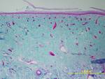
I thought it was stained with a Mallory Trichrome because I know that stains collagen fibres light blue and am sure it is connective tissue but now I'm not sure I'm thinking H&E possibly????
Also, does anyone else agree that this sample is most probably connective tissue from skin? I thought this because of Mast Cells, seems to be rather vascular, appearance of what I think are collagen fibers.... but any ideas from where? I had a crazy idea of maybe eyelid? But I realise Im probably completely wrong.
I'm sorry if this question is very basic but this is my first histology subject and I've been going through textbooks all day and dont seem to be getting anywhere. Thanks for anyone for any help or direction they can give me with this!!!!!!!
microphotographs are attached....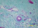

H&E stain will have blue nuclei. so here u have pinkisk nuclei. so I guess not H&E.
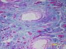
i am not sure if its skin or not. could b skin, need a better magnification of the second pic.
i am not sure if its skin or not. could b skin, need a better magnification of the second pic.
I know H&E stains nuclei blue but Mast Cells are metachromatic so that means they change structures that normally stain blue to red/purple/pink ??? Thats what the textbooks are telling me anyway but I dont always trust what they say (mmm paranoid much?)
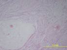
here is a better magnification of second pic.
it seems to b mucosal layer of some region, but still cannot b sure of keratinised epithelia. this region could lead to the skin like in case of lip from mucosa to outer skin or the genitalia.
if u think that there is a lot of mast cells, it is possible.
there r many possibilities for this region, one could b nasal mucosa due to the many blood vessels. it could b lip or something simialr region.
do u have any other pics ?
if u think that there is a lot of mast cells, it is possible.
there r many possibilities for this region, one could b nasal mucosa due to the many blood vessels. it could b lip or something simialr region.
do u have any other pics ?
Do you think the granulated masses are Mast cells? I mean..I know basophils appear similar butttttt mmmmmm they are quite rare and these are quite numerous. Im at a loss. I mean I know its connective tissue but there is no epithelium there...just what appears to be a keraton layer. driving me insane.
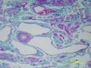
yeah I have more pics also the same tissue with a different stain...


I think the first two stains in this group of pics is H&E (the pinky one) but im just confused because I know H&E is supposed to stain nucleus's blue and everythings damn pink.
About it being mucosa...I cant see any of the layers that are characteristic of a mucosa?? and no epithelium on top or anything...unless the stain is just not bringing it up....
the whole tissue seems to have no organisation to it whatsoever. Just a higgeldy mess of what seems to be adipose (the white blobs), mast cells, collagen, and some cell nucleus's what Im assuming are fibroblasts. but then WHY arent the stupid fibroblasts staining blue in the H&E??
Also Im confused as to what the deal is with the parts of what looks like a mix of pale blue and pink fibres in the first slides.
im going insane hey...
ok, the first 2 pics r H&E, look closely, the nuclei r dark blue and eosin stains others which is pinkish.
the mucosa I suggested earlier can b seen in the pic that u magnified for me. there is the epithelia and i am guessing it to b mucosa.
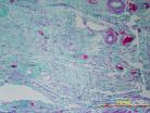
first thing, is there r lots of connective tissue in the pics u have shown. no doubt. i am not sure abt granulated mass, i doubt it. May b some inflammation or infection giving this apperance of inflammatory cells, which is quite possible.
i cannot judge with this magnification abt adipose tissue. Also I have a feeling that there might b muscle fibres as well. of course there r many blood vessels.
do u have higher magnification pics. Do u c any glands - mucous or serous in any sections.
try to look and identify things that u know and then go from there. dont try too hard as u will not get the answer immediately. relax and try to go one by one.
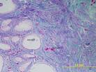
That last pic looked like there are prostate glands on the left, but I could be wrong. I don't think whatever tissue this turns out to be is normal.


Here is what I have so far.....
I'm pretty sure it is loose connective tissue (collagen fibers would suggest this)...but that is as far as I have got. If anyone thinks I am wrong and its dense irregular PLEASE tell me because I'm not 100% confident!!
Ive entertained the thought that it is a MUCOSA...however the top layer you can see looks more to me like a KERATIN layer and I cant really see an Epithelium layer of cells however it IS a low resolution and this might be why...any ideas??? Its very very vascular as well, as you can see.
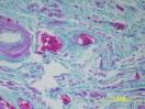
I think the large white circular blobs (for want of a better word) are follicles..possibly ovarian follicles? Not adipocytes because too big for a single fat cell.
At first I thought those massive purple granulated looking things were MAST CELLS but then realised they are a bit too big....Now I'm pretty sure they are blood vessels that have been cut transversely and there are still all the blood cells in them. However Im confused because the other veins and arteries dont seem to have blood cells stuck in them,...agghrrrr!!!!
I still think maybe the smaller granulated masses are either MAST CELLS or another type of GRANULOCYTE..possibly BASOPHILS???
There seem to be a whole bunch of small cells swimming around...now due to it being low resolution Im not quite sure but I figure they are a mixture of FIBROBLASTS and BLOOD CELLS???? any ideas??
Also am tooootally confused as to why in some areas the blue bits become marbled in appearance with a lot of pink. do you think that is just loads and loads of cells? Stupid low magnification cannot see a bloody thing. I want to kill the person who took my photoss!!!!!!!!!!!
THANKS for ANY help at all you can give me....I'm going absolutely insane . I realise Im being very annoying about this its just I have tried so hard and it was due today so its already a day late and yeah im going to end up in a loony bin hallucinating about different types of tissue if it continues on like this...


all organs have an epithelial layer, it only a question of which type of epithelia. u need a better pic of the 1st one to decide on that.
ur sections r highy vascular, so its even more confusin
I think the things u have pointed out in pic 3 & 4 r arteries surrounded by veins.
i doubt it being ovary. there should b lots of follicles which r circular not oval.
try to think of a region which is highly vascular