Immunostaining- opaque layer on fixed cells - Immunocytochemistry (Jul/14/2009 )
Hi Everyone,
I've been having lots of trouble fixing and imaging my immortalized neuronal cell lines. Every time I image it with confocal microscopy or fluorescence microscopy (FITC and Texas Red) an opaque layer of liquid covers areas of my coverslip. There are some areas that do not get covered and the cells are fine (distinct neuronal morphology)...but for the majority of the coverglass the cells are covered in this "layer". I use VECTASHIELD with DAPI mounting media. If anyone could please shed some light onto my problem I would be very very appreciative.
My protocol is basically allowing my cells to uptake FITC conjugated to a molecule and then fixing it. After fixing I stain again for another protein in hopes to colocalize. I use 4% PFA to fix my cells for 20 min at 37degrees celsius and wash many times with PBS.
Thanks!
i think we need a picture for this one
Make sure the vectashield is completely occupying the space under the coverslip. I'm thinking you may be seeing an air interface which is distorting your image, but that wouldn't be opaque, per se. Are you sealing your coverslip to the slide with something that you could be seeing?I always used to use nail polish and that stuff would autoflouresce to the point of it being "opaque".
If you hold up your slide, do you see the opaque area? ... Are you sure that it's not just dust or something? Take a lint-free wipe and wipe it down, perhaps with a touch of methanol to make sure it's not something fixed to it. For a slide to become opaque is kind of strange. Do all your slides do this? Just some? Is this just one or two isolated incidents?
And yes, a picture would be great.
Cheers,
-Carlton
Thanks for the response Carlton. I've tried to wipe the slide surface with ethanol to remove some surface dirt but still having this problem. It is a recurring problem with all of my slides. I'm not sure whether it is the Poly-D-lysine that I use to coat the coverglass with causing this uneven layer or the lack of mounting media. I suspect it could be bubbles where those clear areas are...I don't have an exact picture of what I mean but here are 2 images one of distinct cells "in the bubble" and another one is covered by some kind of liquid under DIC mode on the fluorescence microscope.
What do you think?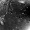
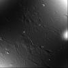
my first impression is that you are missfocusing - it looks like we are seeing glass rather than the tissue/cells inbetween - if the poly D isnt working for you try APES coating the slides - may be smoother
d