Deformed bands in gel - (Jun/16/2015 )
Hi all,
I've been having an issue with my PAGEs lately.
I'm trying to detect a protein of 33 kDa in rat brain samples. These samples are rat brain powder, dissolved in RIPA buffer + protease inhibitors. The problem I'm getting is that there appears to be something in my sample that's disrupting the resolution of the bands in the gel. The bands at the bottom of the gel (which run at about 5 kDa) appear deformed, as if something is 'squashing' the bands. As a consequence of this, the marker is also deformed at that height.
I'm wondering whether the fat content of the samples is too high and may be causing this deformation. During the running of the gel, I can see that along the height of the blue front, something is being pulled down as well that looks like it's working its way through the gel. The result is a smile-effect over the entire length of the gel.
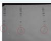
I hope I'm making any sense. Please see the picture attached (it's a bit dark).
Technical details:
- Gradient gel 10 - 20% (Criterion TGX precast), 18 slots midi gel
- Samples contain Laemmli + DTT
- Samples are boiled for 5 minutes at 100 degrees prior to loading
- Loading volume is 28 ul (max 30 ul)
- Protein per slot is 4 ug
- Running is done at 120 Volts, for about 1 to 1,5 hour
I have had such problem before. DNA contamination of your protein sample could do that. Specially if you are extracting with RIPA this happens a lot. Use sonicator or 1 ul of DNAse on your protein sample. Or maybe you were not careful when removing supernatant and accidentally touched the pellet. Next time reduce voltage to 100 V and add up to 20 ug of sample. 4 ug is very low.
Hi Curtis,
thanks for your reply! The reason I only load 4 ug is to reduce background. I started with 20 ug protein per slot and I got incredible smearing in my lanes. The 4 ug seems to be enough to detect my protein (I get bands at the expected height). Our method of preparing protein lysated from the brain homogenates was to simpyl add RIPA buffer and directly use that in the blots. I have a feeling that the samples are too contaminated to run a decent gel (although the problem resides only at the bottom of the gel). I first though that, because they're brain homogenates, maybe the samples were too fatty, but DNA contamination sounds likely as well. I'll see if adding DNase has any effect.
it could be other protein contamination from insoluble cell components. you must centrifuge.
I will also do that!
what is the formulation of your ripa?
if you did not perform a buffer exchange prior to addition of laemmli sample buffer then components of the ripa may be causing the effect you're seeing.
lipids, detergents, salts, etc can all cause these artifacts.
Hi mdfenko,
Thanks for your input! The formulation of the RIPA buffer is: 25mM Tris•HCl pH 7.6, 150mM NaCl, 1% NP-40, 1% sodium deoxycholate, 0.1% SDS. It's a ready-to-use bottle supplied by Pierce.
According to my supervisor and several internet sources, it should be a suitable buffer for Western blotting.
suitable if you 're not interested in the bottom of the gel.
np-40, as well as triton x-100, can displace sds from the proteins and affect migration, but this is not your main problem.
the salt and detergents from your ripa will have an effect on the bottom of your gel. you can avoid this by dialyzing the sample before adding the sample buffer (drop dialysis should suffice, you don't have to dialyze the entire sample, just what you need to load). or, if there are solubility problems, you can dialyze against sample buffer (without tracking dye and glycerol which you can add to the sample prior to loading).
mdfenko: you were right! Instead of dialyzing my samples I first just replaced the RIPA buffer with MilliQ water when diluting my samples, and so far at least the marker looks very nice on the blot! I've only just put on the primary antibody, but I have good faith the band deformation problem has been reduced if not eliminated ") Hooray!
Hooray!