Cloning - Ligation problem - (Apr/01/2015 )
OK, so I have been struggling to clone a 2 kb insert into a 9 kb vector.
Briefly, the steps that I did are as follows:
- insert: amplify using a Hi-Fi Taq from a vector, A-tailed using standard Taq, run PCR product on gel, extract from gel (promega SV wizard), T-A cloned into pCR2.1 TOPO, transform into TOP10, harvest plasmid, digest with EcoRI-HF, cut insert from gel, gel extraction, run on gel & Nanodrop, used for ligation
- vector: cut using EcoRI-HF (then deactivate the enzyme), Nanodrop & run few microlitre on gel to check (there was only one band, so digestion seems complete), Antarctic phosphatase treatment 15 min at 37oC deactivate 70oC 5 min, used for ligation
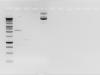
- ligation: I used a molar ratio of 3:1 for insert:vector. I used 3 femtomole (fmol) of insert and 1 fmol of vector in 20 microlitre reaction. Incubation 2 hrs at room temperature. Run on gel the following (lane order): pre-ligation mixture, post-ligation mixture, vector re-ligation (no insert, no phosphatase treatment), uncut vector. I attached the gel image.
Some possible culprits that I suspect:
1. the concentration of insert was actually very low after gel extraction, around 5 ng/microlitre, but if I run on gel around 10 microlitre the band was clear. However, in ligation mixture I put 15 microlitre of insert to achieve the molarity that I needed. This might be not ideal. Should I concentrate it first using ethanol precipitation or phenol-chloroform?
2. I did not see the ligation product in gel (see attached image). At least I should be able to see the insert band or the vector, but it seems that the DNA is disappearing? I did this gel couple of times from different batch of ligation reaction and always get similar results.
The competent cells are OK, it's still new from Invitrogen and tested using other vectors it's working.
What have I done wrong or have I missed something? I need help! Really appreciate your comments and inputs on this.
Cheers,
cahyaprihatna
Have you tried transforming the ligation product? It is not unexpected to see no band corresponding to ligated DNA, and rarely do people run ligation products on a gel.
Your ligation strategy seems excessively complex and difficult. It would be much more straightforward to use PCR to amplify your insert with primers that include two (different) restriction sites (along with a 6 bp 5' overhang to allow the pcr product to be cut. Purify the PCR product, cut with both enzymes. Choose enzymes that can be used in the same buffer and can be heat killed. Then, use similarly designed primers, amplify the vector region you care about, purify and heat kill. Mix, ligate, transform.
This avoids gel purification, which (as you see) has issues, and it avoids phosphatase treatments, which often causes failed ligations. Using two different enzymes allows directional insertion, and avoids easy self-ligation.
Hi,
sometimes I had to purify the digested vector after Antarctic phosphatase treatment to remove the enzyme, to see if this is your critical step,
you should try to re-ligation of vector after phosphatase treatment
Hi,
Thanks for the comments.
@phage434: yes, the strategy that you described is more straightforward and I can try it if I'm still having this problem. However in the past I tried the method to clone something else but it was really hard and I ended up sub-cloned the insert into TA vector. Yes I did transform the ligation mixture despite no visible bands in gel. I now saw some colonies (around 30 colonies). Sadly only one of them is white, the rests are blue. I am growing all of them though, and will check by PCR and digest tomorrow.
@pavoni.ernesto: Yes I also suspect the phosphatase can be the culprit. Will do!