Problems with M13 phage - (Aug/20/2014 )
I am trying to infect E. coli with M13 phage (both purchased from NE Biolabs). I have been following the protocol but still cannot get plaques. I have listed the steps I have been following. I would appreciate any feedback. I am an undergrad, who took over a grad students project, and I have very little experience with microbiology. E. coli is kept at -80C and M13 at -20C.
1. Use frozen E. coli (Biolabs) to plate for single colonies.
2. Add single colony to LB broth, grow to approx OD600=0.4.
3. Dilute M13 (Biolabs) phage of 1 x 10^13 pfu/ml. (I have tried both PBS and LB broth, which Biolabs suggested)
Use microfuge tubes - Add 990 microliters of LB to 10 microliters of M13 to get 1 x 10^11 pfu
Then 100 microliters of that to 900 microliters of LB - 1 x 10^10......and cont until I get to 10^1.
4. LB Agar plates are warmed at 37C for an hour.
5. Top agar is prepared and kept between 45-50C.
6. Mix E. coli and M13 in microfuge tube (by flicking the tube). I have used various quanities, such as
1 ml E. coli : 50 microliters M13,
200 microliters E. Coli : 10 microliters M13 (Biolabs suggest in protocol)
Wait 5 min at RM temp.
7. Add E. coli/M13 mix to 3 ml of warm top agar. Vortex. Pour on LB plates. Allow to set. Invert and store at 37C overnight.
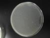
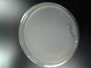
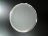
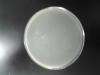
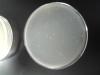
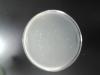
I have used to 10^1 through 10^7 m13 dilutions and so far my plates either look like an overgrown lawn of bacteria or dots of bacteria all over the plate. I have no clear areas that look like plaque. I would love to hear from anyone. Thank you
It is possible that the phage has gone off as I thought it was normally stored at -80, but it has been a loooooong time since I last worked with phage.
However, is there a trend in the dilutions where you see the bacterial "dots" all over the plate? If so, which dilutions show this? If these seem to be dilutions with high titre, then it is very likely that you do have functional phage, as it is killing the majority of the monolayer, and what is happening is that you just haven't noticed the plaques.
Phage plaques can be quite small and easy to miss. There are two techniques that can help you see plaques, one is to hold the plate against a dark background and look for clear patches, and the other is to hold it up to the light. A third that sometimes works if you have a lawn is to try and reflect light off the plate surface such that you can see irregularities in the surface, small holes are often plaques.
Thank you for your reply. The dilutions of 10^5 - 10^7 were just lawns of bacteria. The 10 - 10^4 were spotted. The problem is I also put a control of bacteria alone in that batch and it was spotted also. I held them to the light but not dark background. I will try that next. I have attached the pics but compared to the pictures I have seen of plaques, it didn't look right. The student that I took over for, left me no photos of his one successful plaque growth, so I have just been comparing to pictures I have seen on the internet.
Looks like you need to work on your pouring technique then. I think it would be best if you ignored the density thing and just went with an overnight culture for the propagation step.
You should be able to add the phage and bacteria directly to the top-agar, mix by inversion and pour quickly. Also check that the strain of E coli that you are using is permissive for M13 phage - not all are. You probably want something like JM101 or derivatives.
I have one last question. First, I am using the E. coli ER2738, that came with the M13. and am following the M13 Titer Protocol that I found on New England Biolabs website. The 4th step of their protocol is what I am having a hard time understanding, it is as follows:
Prepare dilutions using 10 to 10^3-fold serial dilutions of phage in LB; 1 ml final volumes are convenient. Suggested dilution ranges: for amplified phage stocks and infected culture supernatants, 10^8 - 10^11; for unamplified panning eluates , 10^1 - 10^4.
The M13 comes as 1 x 10^13 pfu/ml. Maybe I am confused about the serial dilutions. If I dilute it as I stated in #3 of my first post then for example my 10^9 dilution would actually be 1 x 10^4 pfu/ml. All I am trying to do is determine the viral titer. I will be doing an experiment on this virus and after I want to compare to see if less virus grows.
Thank you again
https://www.neb.com/protocols/2014/05/08/m13-titer-protocol
So it looks like the NEB protocol is suggesting doing somewhere in the range of 10-1000 fold dilutions. You look like you have been doing 10 and 100 fold dilutions, so your 10^-9 dilution would indeed be 10^4 pfu/ml.
It seems to me that you probably need to do some dilutions inbetween 10^-4 and 10^-5, perhaps two-fold in this range. Note that 10^4 pfu/ml is 10,000 pfu - you would expect to see 10,000 plaques on a plate if you plated 1 ml. You definitely can't count 10,000 plaques on a plate. Usual range is 30-300 (upper number can be increased, but don't go below 30 for statistical reasons).