MIN6 Cell Culture Contamination Risk - (Aug/12/2014 )
Hello everyone,
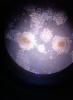
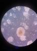
I am working with MIN6 cells which are adherent, pancreatic beta cells isolated from mouse. In passage 3, i saw some clusters that i cannot identify whether they are bunch of cells or contamination. They looked like neuron-like first, now i cannot say what they look like.
Cell media is composed of DMEM, L-glutamine, b-mercaptoethanol, pen/strep, FBS
I have never experienced a contamination in cell culture so i would be very happy if you can help me out. If you have some kind of list about understanding contamination, it would be also helpful. I attached two photos that i had hard time to took. Thanks in advance!
Fatih
Hi Fatih,
To me, this doesn't look like contamination. I'd say the cells are growing in clusters and aren't forming a monolayer but instead are growing 'up' (or rather, forming balls of cells that are not two-dimensional but three-dimensional). I can't say what this means for the health of your cultures, but what you could try next time to prevent this is leaving the cells on Trypsine for just a while longer while passaging the cells. In addition, you can try to detach the cells from each other during passaging by pipetting your cell suspension up and down a lot and then looking under the microscope to see whether you have a single-cell suspension, before you transfer a portion to a new flask.
The reason I believe these clusters to be cells and not contamination, is because I see very similar structures form when culturing rat pheochromocytoma cells, which are notoriously 'sticky' and cluster like crazy. Very large clusters, like this one, are dense in cells and thus get this 'orangy' center. But in your photo's I can also see smaller clusters form (in the second photo, at the upper edge and at the left edge I can see smaller balls of cells).
Be aware that if these clusters get too big, the cells in the center of the cluster are possibly deprived from nutrients, as they're no longer in contact with the medium. As far as I know, very large clusters can detach from the plate and start to float. If you want to prevent this from happening, try following my instructions above ")
As far as recognizing contamination goes: a distinction can be made between a bacterial, fungal, viral, mycoplasma, yeast or cross-contamination. Depending on the type of contamination, it can be either pretty obvious what's going on or very hard to notice; a bacterial infection is usually pretty easy to notice just by looking at the medium color and clearness and viewing the cells under the microscope. A viral infection can be very difficult to spot, I've been told. A mycoplasma infection can only be detected using mycoplasma-detection kits or assays, as these cells are too small to see.
The internet is full of tools and tips on how to spot and prevent infection. Do some research, I'm sure you'll find a lot of information.