Cell culture contamination ? - (Dec/10/2013 )
Greetings,
We have had something going on in our cell cultures lately and I have a hard time determining what it is. We are culturing breast cancer cell lines (with antibiotics in the media in routine).
It first started with a decrease in the cell proliferation rate. I would seed 2 millions cells in a flask and end up with more or less the same number few days later. Even though the flask looked confluent after harvesting and counting the net increase was almost zero.
I suspected a contamination, changed all media, cleaned everything thouroughly, stopped using the waterbath and started back from frozen stocks but still have the same problem.
The media's pH or turbidity never changed and I didn't see anything at high magnification on the microscope, so I ruled out bacteria, yeast and fungi.
Mycoplasma infection was fitting the poor proliferation rate (these cells are supposed to be highly proliferative) so I started treatment with BM cyclin for mycoplasma and sent the cells for testing. The test came back negative but the cells are still behaving the same. Then I noticed that if I look at the flask at high magnification after harvesting the cells from it, I see some clumps of dead cells, together with cell debris and a lot of what seems to be cells "skeleton" or cells emptied from their content and little things moving around the cells remnants. I only see that on the flask after the cells have been harvested and never on the flask before harvesting the cells. Could it be intracellular bacteria that are released in the media from dead/contaminated cells upon harvesting? I incubated the cells in antibiotics free media but so far there have been no change in the media's pH or turbidity and nothing can be seen on the scope (but it's just been 24 hours).
I would love to get your feedbacks, since I am running out of ideas and don't know how to take it from there. I would love to test a different antibiotics combo (other than pen/strep) and was wondering what you would recommend.
Thanks in advance for your help!
It could be a number of things. Could you post a photo of the cells as they appear down the microscope, and a photo of the "skeletons".
Are these a primary/non immortal line? Has your batch of FBS changed recently?
They are immortalized cells. We haven't changed our FBS source (same ref, same lot number).
I don't have a pic of the "skeletons" but will try to get one next time I harvest the cells which is the only time I see them floating in the flask once I remove the cell suspension.
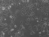
I am attaching a pic of the cells. I am wondering what are the punctuation that we see in the cells (they don't move and are refringent)?
The media in the flask where the cells grow in antibiotic free media is still clear after 48 hours, although slightly orange (but the cells are approching confluency). No obvious sign of infection is seen upon inspection of the cells. The cells have the same aspect as the ones growing in media + pen/strep.
Thanks for your feedbacks!
Best,
The cells look fine. The refringent dots in the nuclei are nucleoli, dots in the cytoplasm are probably endoplasmic reticuluum or golgi apparatus, but could also be cell-associated bacteria (unlikely unless they proliferate).
Do you prepare your own medium (i.e. do you buy pre-made medium or not)? Are you adding any additives like L-glutamine to the medium? Do you have phenol-red in your medium (breast cells are usually estrogen senstive, phenol red is an estrogen analogue...internalization of the estrogen receptor will slow growth rates)? Have you checked the CO2 concentration of the incubator? Does the medium appear to be buffering correctly in the incubator (incubate some with no cells)?
We buy pre-made medium (with phenol red and glutamax) and add non essential amino acid, FBS and pen/strep (everything is aliquoted). I've tried adding beta estradiol since it is supposed to increase cell proliferation on these cells (they are estrogen receptor positive) but it didn't make a difference.
Haven't physically checked the CO2 concentration in the incubator, the probe indicates the expected CO2%. Haven't check the buffering capacity of medium with no cells. Will do that next! Thanks for the tips!
That's unusual, ER+ cells (e.g. MCF7) usually have a slower growth rate if the receptor is activated and internalized.
A lot of the breast lines are supposed to be grown with insulin too. Though I must admit that I haven't had any problems with the MCF group with regards to this.