help with evaluating kidney pathological changes - (Aug/20/2013 )
Hi everyone,
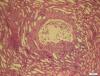
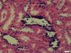
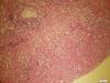
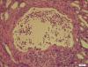
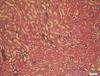
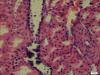
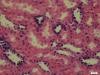
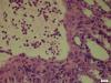
We have some renal biopsies of rats in a toxicologic pathology test, and we found cysts in the kidneys. Someone considered that it probably caused by calcification.
I've never worked with rats. If anyone could help me out and tell me what is causing the tissue to have lesions in it I'd greatly appreciate it!
Thanks in advance.


Hi everyone,
We have some renal biopsies of rats in a toxicologic pathology test, and we found cysts in the kidneys. Someone considered that it probably caused by calcification.
I've never worked with rats. If anyone could help me out and tell me what is causing the tissue to have lesions in it I'd greatly appreciate it!
Thanks in advance.


It would be helpful if you told us what we were looking at (magnification, part of the kidney, etc.).
fhlz2013 on Tue Aug 20 10:46:02 2013 said:
Hi everyone,
We have some renal biopsies of rats in a toxicologic pathology test, and we found cysts in the kidneys. Someone considered that it probably caused by calcification.
I've never worked with rats. If anyone could help me out and tell me what is causing the tissue to have lesions in it I'd greatly appreciate it!
Thanks in advance.