Distorted bands from human colon samples - (Jul/23/2013 )
I have been running western blots for some time now and I am having difficulty running some human colon samples that perhaps I could get advice on. In brief my protocol is as follows:
To 0.12g of colon tissue, I add 1ml of RIPA (0.96g Tris, 3.5g NaCL, 0.24g EDTA, 0.3g EGTA, 4g Na Doxycholate, 4ml Triton-X 100, 4ml 10% SDS, made up to 400ml with dH2). I leave the tissue in RIPA for 10 mins on ice, after which I homogenise using a rotor homogeniser for 10 secs. The slurry is then passed through a 25G needle, and put into a sonicating ice-bath for 1min and then spun at 12000G for 10mins at +4. The supernatant is added to x4 LDS buffer (invitrogen) + denaturing solution (invitrogen) and heated at 90 degrees for 10 mins. I typically load 8ug of protein into a Bolt 4-12% bis-tris gel and run at 165V (recommended by invitrogen) in 1x MES buffer (invitrogen) for 40 mins.
The problem I am having is that the samples are not running flat. The normal musocal samples look like typical bands when ponceau S is added but the cancer samples tend to look condensed and distorted making comparison impossible. I have enclosed an image that shows what I am seeing. The blue dye front is fairly representative of the sample issue I am having - some look normal, others condensed.
I have tried running the gel at a lower voltage for longer but the problem remains. I am beginning to wonder if DNA is playing a part in this. Any advice would be most welcome.
Regards
Dr Ross Hamilton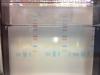
I would guess there is a problem with your salt content somehow. I have seen similar things in a colleague's BN PAGE gels, but can't for the life of me remember how he solved it.
i don't think it's dna and i'm a little puzzled as to why it effects the cancer samples and not the normal samples, but...
some of the things that could be causing your problem are salt (as mentioned by bob1), triton from the extraction buffer (may prevent lds from binding to the protein), lipid and/or carbohydrate contamination.
you may want to try precipitating the protein away from the extraction medium with tca before preparing it for electrophoresis.