Unknown mitochondrial morphology? - (May/30/2013 )
We have been trying to figure out what these structures are in our electron micrographs of mouse muscle (longitudinal images). The second image is from mice fed a high fat content diet. The structures don't appear to be fat droplets though, as other EM images of fat droplets appear darker and totally smooth. That could just be different staining/contrast techniques though. It doesn't appear to be autophagy of mitochondria either as there is nothing "surrounding them". Could it be some sectioning artifact? It only happened to high fat diet though. It looks like the mitochondria are swollen or something. You can see what looks like cristae in them (and in other images not shown). I can't find any other reference papers or images with the same structures though so I'm having trouble confirming it. Has anyone seen these before?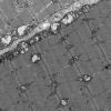
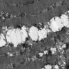
They certainly look unusual, I would have said that perhaps there was brown fat in these, rather than white fat.
I also just found a paper in Mitochondrion, vol13 2013 - have a look at fig 5.:
Multiple Symmetrical Lipomatosis—A mitochondrial disorder of brown fat
Plummer, Spring, Marotta, Chin, Taylor, Sharpe, Athanasou, Thyagarajan, Berkovic.