Transcription/Translation reaction not resolving - (Mar/15/2013 )
I have done overnight in vitro transcription/translation reactions that produce a 200 Kd protein that can not self-cleave (Lane D) or can self-cleave (Lane E). I have attached the gel image.
In lane D (can not cleave itself) I do see a strong band, albeit at a lower MW then expected, but I think it is right.
In lane E I should observe cleaved proteins (5-6) and a few intermediates, instead I get a smear with only one band visible (about 1/3 down from the top)
I ran a 4-20% HEPES gel from Pierce. I use SDS-HEPES buffer. I have treated all samples with 2X beta mercaptoethanol SDS loading buffer, heated at 70C for 10 minutes, and ran for 2 hrs at 30 milliamps. I exposed for 90 minutes (overnight does not show additional bands)
Can anybody explain the smears? I should be seeing bands from 20-60 Kd in lane E (possibly intermediates at higher MW).
I used the TnT Quick Coupled transcription/translation kit from Promega. The lysate is very viscous and is difficult to run on gels. If anybody has experience with the Promega TnT kit, please let me know.
In short, I really need to improve the quality of these gels with better resolution. My constructs have been sequenced and they should be fine for the TnT reaction. There should be one band in lane D, and around 6 bands in lane E. I am hoping the additional smears in lane E contain the desired proteins and I am simply running the gels incorrectly, somehow.
Thanks for any advice.
JM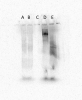
It is entirely possible that the cleavage depends on correct folding of the protein, which may not happen in vitro, it could also depend on a post-tranlational modification such as phosphorylation, that also probably wouldn't happen in vitro.
bob1 on Sat Mar 16 00:48:53 2013 said:
It is entirely possible that the cleavage depends on correct folding of the protein, which may not happen in vitro, it could also depend on a post-tranlational modification such as phosphorylation, that also probably wouldn't happen in vitro.
I agree these could be factors, but this has been done by a couple groups in the literature, and I simply need to repeat as a portion of my work before I can move forward. I have followed their instructions on the TnT reactions, and I think the problem lies within the gel running. Their methods didnt contain the fine details of their electrophoresis, it just read "resolved translation products by SDS-PAGE."
what's in your extraction buffer? salt can cause what you are seeing.
so can detergents. if you have non-ionic detergent, like triton or nonidet, it can displace sds from the protein.
mdfenko on Mon Mar 18 18:13:35 2013 said:
what's in your extraction buffer? salt can cause what you are seeing.
so can detergents. if you have non-ionic detergent, like triton or nonidet, it can displace sds from the protein.
I dont do an extraction. This is a commercial rabit reticulocyte lysate prep. I just add DNA plasmid and the reaction does transcription followed by translation. I then take ~5uL of the reaction and mix with SDS sample buffer and run on a gel.
I did add PCR enhancer to the reaction since it was reported that Mg2+ is important for proteolysis. I could try reducing the amount I add.
JM
the lysate probably contains a physiological concentration of salt. this can cause problems with page.
you can eliminate the salt by drop dialysis or you might be able to ameliorate the effect by dilution of the sample prior to addition of the sample buffer.
Thanks for the tips. I will post what works when that time comes (hopefully).
JM
I figured it out. By loading 1/3 the amount of translation reaction and using LDS sample buffer with NuPAGE gels, the gels look very nice. The biggest thing was the overloading. With a TnT reaction, I assume there are a lot of components that interfere with resolving a gel.