bands position in agarose gel - (Dec/29/2012 )
This is a silly question but me not being a molecular biology person am unable to understand this.
we perform plasmid extraction using miniprep kit (alkaline lysis method).
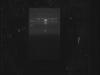
Attached please find the gel. All the samples are of the same clone (lane 5 is hind III marker) but analysis perfomed on different time. Our results should look like the one in lane 1, however we get bands in higher mol wt. Can someone explain this and what we can do to overcome this.
we use TAE buffer during the run and add ethidium bromide during the agarose gel preparation only.
 .
.
thanks in anticipation.
Think about the forms plasmids can have.
i do know that supercoiled plasmid moves faster and niked relaxed plasmid will move slower, however we perform simple plasmid extraction using kit. so how can the plasmid be nicked. what is then the sureshot way to get proper results as with this results some expect the plasmid is different.
thanks
The forms are supercoiled, nicked (circular) and linear. Most extractions will have all 3 forms (there are actually a few more, but you are unlikely to come across those) unless something has gone wrong, in which case the DNA will mostly be linear.
The forms are generated both in the bacteria and by the extraction (2nd step is alkaline lysis) where the DNA is actually degraded by the alkalinity of the solution. The amount of relaxed circular is also dependent on the removal of protein during the extraction process.
Have you checked the purity of the sample by NanoDrop or related method? (260/280 nm)
If your miniprep failed to remove proteins effectively, you might be clogging your lanes and so your sample is getting into the lane late. If you look at the well up top, you can see there is some build-up right where the lane starts.