Please help with odd bands and smears on Western blot - (Oct/18/2012 )
I am having trouble with my Westerns. We are looking at beta-catenin and using beta-actin as a loading control. My beta-catenin looked smeared. This is the first time this has ever happened. Also some of my beta-actin bands look scrunched up instead of having nice straight bands. Any thoughts?
1. How did you prepare your cell/tissue lysate?
2. SDS-PAGE running time/settings?
3. Did you block your membrane before primary + secondary staining? With what & how long?
4. Primary/secondary antibody dilutions + incubation times?
5. Did you probe the same membrane twice (first for b catenin and/or b actin followed by b actin and/or b catenin)?
science noob on Thu Oct 18 20:44:17 2012 said:
1. How did you prepare your cell/tissue lysate?
I lysed BMDC in 75 uL of RIPA buffer with protease and phosphatase inhibitors added. I then froze them at -80.
2. SDS-PAGE running time/settings?
Running time was about 50 minutes on 175 mV.
3. Did you block your membrane before primary + secondary staining? With what & how long?
Membrane was blocked with 5% NFDM for 1 hour. Primary was left overnight, washed 3 times with TBST and then the secondary was added for 1 hour.
4. Primary/secondary antibody dilutions + incubation times?
Primary dilution - 1:1000 for both antibodies overnight at 4 degrees.
Secondary dilution - 1:3000 for one hour at room temp.
These parameters are all suggested by the antibody company (Cell Signaling).
5. Did you probe the same membrane twice (first for b catenin and/or b actin followed by b actin and/or b catenin)?
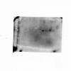
I probed twice. First for b-cat and then for b-actin. The attached images are of the same membrane.
It looks like either your gel or running buffer isn't prepared properly. Try remaking the running buffer. You should also use less secondary - the presence of the ladder as a negative band indicates high background, which is usually due to the secondary being at too high a concentration.
Despite the manufacturer's recommendations, antibodies almost always need to be titrated for best use as the signal depends on a whole bunch of factors including the blocking conditions, membrane type, amount of protein loaded, incubation time...
Yup, I agree with bob1 that secondary antibody dilution can be increased to 1:5000-1:10,000 ( which would explain the background, 'smear' and nagative band in the ladder lane as mentioned my bob1)
The scrunched band seems like a loading/running problem. This normally happens when your pipette tip pokes into the gel when loading, giving it a 'rounded' leading front.
Did you prepare all the samples (all lanes) in the same lysis buffer on the same day?
Did you add a reducing agent (DTT or b-mercaptoethanol) in your cell lysis buffer and boil lysates before running it on the SDS-PAGE?