colony cracking for screening of positive clones - (Oct/17/2012 )
Hi,
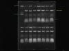
I have tried the colony cracking method to screen for postive clones. The pYES260 plasmid size is 5933bp.I digested it with BamHI (1 site) and filled-in to create blunt end plasmid. My PCR insert is about 2kB. After electroporation and plating, I extracted the plasmid using colony cracking. However I am not sure which band represented my plasmid.
The first lane on the upper left is the pYES plasmid while other lane are my clones.
Is what I assumed correct?
<*>The pink line is the band for genomic DNA
<*>The blue line is where two bands were observed would be the RNA (Is the faint band across the lane is RNA?)
<*>The purple line is where the proteins were.
<*>The yellow line is the PLASMID.
Another thing is, when I extracted the plasmid, it yielded the three bands. I am not sure of which size should I refer to compare with the clones.
Can anyone have a look at the gel picture attached?
-Helena Kyle-
Thank u 
<*>The pink line is the band for genomic DNA
Probably yes. This is DNA that is still trapped in the well, so likely to be genomic.
<*>The blue line is where two bands were observed would be the RNA (Is the faint band across the lane is RNA?)
I would say nope. I very much doubt that RNA would come out nice clean bands like this, I would say these are more likely your plasmid in two separate conformations.
The RNA is likely to be the smear at the bottom that you have marked with the purple line.
<*>The purple line is where the proteins were.
I wouldn't expect to see protein at all with AGE....(and as I said, my guess is this is your RNA)
<*>The yellow line is the PLASMID.
agreed
As for which bands to compare, I can't tell you as I've not used this method before... sorry
Thanks very much for your help, leelee 