【help】What could happen to this SDS-PAGE ? - (Apr/06/2012 )
I use the 4-20% gradient gel from Pierce. The picture shows the Sypro Ruby staining for whole protein after running. It was generated by a scanner. Naked eye observation on a transilluminator is very similar.
What confuse me is that Ican see the bands, but they are not distinct from the background and difficult yo be compared. The edge of each gel lane has great stainning and appears like smear. 
I thought I had a high concerntration of Na+ and K+ in the solution ( both are about ~500mM) ,which may cause the protein aggregate. But after I tried to use choloroform to desalt and purify the protein, I found it gave little help 
Anyone had any idea about this problem?
P.S.: I do not know what is the two big dots of strong signal at the bottom .....
Thank you very much!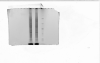
probelms with proteases/proteolysis? this would explain the grey background and the big dots of protein debris/peptides at the bottom
use protease inhibitors at each step of preparation (even in Laemmli buffer)