Trypan Blue Exclusion Test - (Feb/15/2012 )
Hi all!
I am just using this assay for the first time.
I need to count cytotoxicity from primary rat hepatocytes.
I can easily detect the live cells as there is no staining.
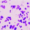
However, in case of dead cells, different patterns are seen.
Some are irregularly shaped, some are round with dark blue spot, some are light blue but no round spot.
I am attaching one image that I found from Internet.
Hi,
Basically, Trypan Blue is only incorporate in dead / dying cells. However, notice that trypan blue is toxic, so if you wait too long you can observed some "funny" cells. So if you look at your cells quickly, blue is dead and others alive. The shape of you dying cells can be explain by the type of death : apoptotic or necrotic, I guess dark blue spot are for late apoptotic cells but not sure
If you want to assess apoptotic or necrotic cells, you should use annexin V protocol, this could clarify the situation.
Hope this help, good luck
Thanks ThibautEBV for the suggession!