Moving "rods" in human PBMC culture? - (Dec/02/2011 )
Hello everybody
in our lab we have to isolate PBMCs from human blood using Ficoll and culture them with different stimuli, afterwards. Recently, we found weird looking "rods" in between the cells which are moving but we do not know what these rods might be. We thought about platelets but we do not know for sure. I made a pictuire of our cells, and I marked the "rods" (at least 3 of them) so that you know what I mean. I'd really appreciate it if you can help us! Thanks in advance,
Nono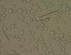
Way too big to be platelets.
Looks like some pseudohyphae at the 6 o clock position, kinda of hard to tell but I'm leaning towards fungus,
Thanks for the reply.
Well, if it's not platelets we don't have any idea what it could be. And fungus... We don't know were it might come from.
Yesterday we tested our methods again using my blood. The blood was taken in a sterile way and also the isolation proceeded in a evry and careful manner. We desinfected everything between every step, we even cleaned/purified/desinfected the incubator and the laminar flow.
But even thenyou could already see these rods 2 hours after plating them in a 48 well plate. We even plated the solutions we used (FBS, culture medium, ack lysis, PBS etc). We are really desperate because the cultures are very important ;(
If you are working with patients, perhaps the 'rods' are bacteria present in the patient's blood itself? If you are working with healthy donors though this possibility is out, since I don't see how a donor could be fine and feeling normal while having sesis....!
On the other hand, have you observed the motion of these rods? Do the rods only appear after you treat with stimulus or right from the start? Because I extract PBMCs and infect them with various bacteria for my own project, and I have noticed that after infection of the PBMCs, rod or sickle shaped cells appear, which upon looking at for longer, are actually the phagocytic cells performing ameboid movement. So I see the sickle/rod shaped cells, and then when I keep looking at it for ~5-10 seconds, it would spread out again and come back into focus as a fully circular cell, and this would repeat.
Hope you figure out a solution though, and if you do, please keep us posted as well!
Well, we do use blood from children that have asthma but my blood was taken as a control and I don't have any asthmatical symptoms or anything else. So an infection in the blood itself can be more or less excluded.
We stimulate the PBMCs with following stimuli:
1. unstimulated
2. Phytohaemagglutinin (PHA-M)
3. Rhinovirus
4. HeLa cells
5. R848
6. LPS
7. Poly I:C
8. endotoxin-free bacterial DNA (E.Coli)
9. Zymosan
We observe these rods in every condition even in the unstimulated culture, but not with the PHA-M-stimulated cells.
The movement is no migration from point A to B (at least not evry much) but more like a kind of shivering and coiling up. Sometimes, when I observe one of those rod for a longer time it gets kind of blurry and then takes a spherical form or it elongates.
Thanks, I also hope that we can figure out what it might be. Any other ideas would be really helpful!
I'm having the exact same problem here...
I've repeated many times, washed every thing, changed all medias, ficoll, pipets etc.
I have abosolutely no idea were this can come from or what can it be. Anyone can help me?
Nono and SD1,
Did you find the answer? I have the same problem :( Could you please help me?
they could be red blood cells that have come through your prep.
MEV on Tue Sep 16 12:47:10 2014 said:
Nono and SD1,
Did you find the answer? I have the same problem
Could you please help me?
Hello
I have the same problem in my PBMC culture, did you find the answer or have any other idea? Please I need help too. 