No bands in cytosolic extraction, nice bands in nuclear extraction--see pics - (Jul/22/2010 )
Hi everyone,
I am extracting protein from endothelial cells using a cytoplasmic/nuclear extraction kit from Cayman Chemical (https://www.caymanchem.com/app/template/Product.vm/catalog/10009277/a/z). The protocol provided by the manufacturer is followed exactly, and our Bradford assays show that protein is present in both extractions (about 1-2 mg/mL in cytosolic, about 0.5-1 mg/mL in nuclear).
For SDS-PAGE, we use Invitrogen Bis-Tris 4-12% gels as well as their LDS sample buffer and reducing agent. Upon completion of electrophoreis, we transfer to a nitrocellulose membrane. We then complete the western blot by adding an antibody for the glucocorticoid receptor (GR).
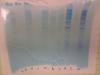
However, there is an issue that has occurred ever since I started SDS-PAGE/western blots last month. After staining with Coomassie Blue, I am consistently getting very little to no bands in my cytosolic protein lanes, but many bands in my nuclear protein lanes (see attached picture). Furthermore, when I blot for actin (housekeeping gene)in my western blots, it always appears in the nuclear lanes! The cytosolic lanes either have no actin bands whatsoever, or a very faint band...this is very unusual (see attached blot). Regardless, I still get GR bands in our control cytosolic lanes, and after stimulation with its steroid agonist dexamethasone, the bands appear in the nucleus.
So, even though my protein of interest is appearing in the proper extraction groups, I feel that the issue with the bands on the gel and actin should not be a big issue. But, it is certainly bugging me and I have yet to determine what could be the problem.
Your help would be greatly appreciated!
Pictures: An image of the dryed gel is attached. I labeled cytoplasmic lanes with a "C" and nuclear lanes with an "N". The ladder lane is marked with "L". Sorry for the image quality--I took it on a cell phone camera. The other blot image shows GR (top bands, order is cyto nuc cyto nuc...ignore the right side of the blot); notice actin (lower bands) is present in only the nuclear lanes.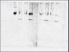
Hi there,
I am wondering if you load equal amount of nuclear and cytoplasmic protein?