Is this a contamination or what? Please help me ! - (Apr/08/2010 )
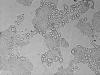
hi, I had a cell culture problem recently. The immortalized MDCKII cells did not function well and if you look from the microspopy, there are some bubble like stuff. Does any one know the reason? Is this a contamination?
I attached the picture below.Please take a look. Any comments will be highly appreciated.
thank you !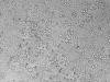
Doesn't look like contamination to me. Look more like vacuoles. How was the media - any change in colour, maybe your CO2 levels were off or you had made them too confluent previously or they're an high passage number?
Just some thoughts, but doesn't look like contamination.
Its a little hard to tell but it looks to me like your cells are over confluent in the areas where they are growing, leading to them senescing and dying, which is the cause of the bubble like appearance (vacuolation during senescence).
Thank you for your kind reply. The media looks normal: clear and no change in color. The passage number is quite low, it's about 6 or 7th generation.
I also suspect for the over confluence issue. The tricky part here is that I need to do the directional flux study which requires the 100% confluence of the cells on the transmembrane. Should I add any more nutrition to the media to make the cells happy if this is the case?
LostintheLab on Apr 8 2010, 05:23 PM said:
Just some thoughts, but doesn't look like contamination.
Are you using the media for MDCKII cells (the one that the ASCB recommends etc)?
I've not cultured these cells to be sure of how to improve their culturing, maybe adjusting the serum levels might help, but I would try that on a smaller culture to see if its worth it or not.
LostintheLab on Apr 9 2010, 01:23 AM said:
Just some thoughts, but doesn't look like contamination.
Your spot on lostintheLab.......vacuoles they be. This sometimes happens if you over trypsinise your cells. It also happens when you tranfect some cell types.....as bob1 rightly states, vacuoles appear as part of cellular senescence. It is also a thing you see with some types of Primary Endothelial cell.
Kindest regards
Rhombus
rhombus on Apr 9 2010, 02:53 AM said:
LostintheLab on Apr 9 2010, 01:23 AM said:
Just some thoughts, but doesn't look like contamination.
Your spot on lostintheLab.......vacuoles they be. This sometimes happens if you over trypsinise your cells. It also happens when you tranfect some cell types.....as bob1 rightly states, vacuoles appear as part of cellular senescence. It is also a thing you see with some types of Primary Endothelial cell.
Kindest regards
Rhombus
Agree....The HEK293 I transfected sometimes, or more precisely, quite often have this scene. I also do not know why vacuolization happened even I just wash them with PBS or refresh medium.....
It does look like vacoles. I always got vacuoles in my MDCK cells growth from ATCC but it looks much smaller than yours and more like in the cells. but yours looks like within the space of cells (I am not quite sure about that....). But after I remove the Amphotericin B from the media, the cell grow fine. Did you put any fungizone in your media?
lilyac on Apr 8 2010, 06:32 PM said:
I also suspect for the over confluence issue. The tricky part here is that I need to do the directional flux study which requires the 100% confluence of the cells on the transmembrane. Should I add any more nutrition to the media to make the cells happy if this is the case?
LostintheLab on Apr 8 2010, 05:23 PM said:
Just some thoughts, but doesn't look like contamination.