Loading Equal protein concentrations - Does it make a difference in simple overexpression checking? (Mar/25/2010 )
Hey guys, so this is my fourth post on this forum, recieveing some good replies to my questions, so happy.
My question is, does it make a massive difference to load te exact same concentration of total cell lysate(Bradford assay verfied concentration) to the wells when comparing overexpression between an induced BL21 culture and an uninduced BL21 culture?
Because i did the same protocol on both samples, i.e induced/uninduced at same OD, bugbustered at same time and volumes etc, and added the same volume into the wells. However, the bradford showed my control was 50 mg/ml and 20mg/ml was the induced sample(only did this after i loaded samples and got the gel). I loaded 10 ul of each sample into SDS-page. Now, I think i see a nice juicy overexpression band at my size of the protein, but I also see a nice thicky juicy overexpression band in my control?! So I assumed my expression failed. However, I noticed the control lane has much darker stain and the bands are much better stained. Possible that its a false-positive control and in realiy there is no thick juciy overexpression band in the control, but just looks like it because it overloaded in relation to the induced lane?
Sharky on Mar 25 2010, 09:07 AM said:
My question is, does it make a massive difference to load te exact same concentration of total cell lysate(Bradford assay verfied concentration) to the wells when comparing overexpression between an induced BL21 culture and an uninduced BL21 culture?
Because i did the same protocol on both samples, i.e induced/uninduced at same OD, bugbustered at same time and volumes etc, and added the same volume into the wells. However, the bradford showed my control was 50 mg/ml and 20mg/ml was the induced sample(only did this after i loaded samples and got the gel). I loaded 10 ul of each sample into SDS-page. Now, I think i see a nice juicy overexpression band at my size of the protein, but I also see a nice thicky juicy overexpression band in my control?! So I assumed my expression failed. However, I noticed the control lane has much darker stain and the bands are much better stained. Possible that its a false-positive control and in realiy there is no thick juciy overexpression band in the control, but just looks like it because it overloaded in relation to the induced lane?
It might make a massive difference. It might make a little difference. If it makes any difference, you should do what you can to eliminate that difference. If you're looking at expression/overexpression, you can't load two different amounts of protein, because the difference in signal could be because more target rather than more expression. Your controls are at least 2x more concentrated, so the target band will be twice as intense as it would have been if you loaded closer to 200 ug. Your "fat juicy" bands might also be saturated signals, so you should do serial dilutions to find out how much you actually have. Once the signals are saturated, you can't tell a difference even if in reality there's a big difference.
One other thing is induction. Is this with IPTG-Lac induction? If so, lac is frequently leaky, so you'll get expression without induction. Induction just increases the expression.
So, I would suggest starting out loading 200 ug (or less... that's a ton of protein) and then do 1/5 or 1/10 serial dilutions to see where the control signal dilutes out relative to the induced. As I've stated in another western thread, using a reference protein is useful for such an experiment, particularly if you plan on using any of this info in a publication (or thesis/dissertation).
Ah, I see what you mean. Very concise explanation, thanks fish!
Further questions:
1) What classifies a good expression on a gel? Just an increasing intensity band of your protein size? How can you compare the expression of the same gene in two different cell-types using these bands? Any easy way? I dont have a densitometer in the lab.
2) What do you think is the best protein concentration to load on a Nupage 4-12% Bis-Tris gel to see optimum resolution and seperation?
On the invitrogen website(Picture attached) it says total sample volume should not exceed 15 ul, and maximum load for optimum resolution is 0.5 ug/ coomasie stained band? I dont understand what the latter part means, how does that apply to a lysate?
3) Doing a bradford assay before loading to the cell and adjusting the concentration of protein added to sample buffer for my control and induced to make sure they are equal will gaurantee that I can reliably compare the two lanes?
Thanks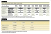
Sharky on Mar 25 2010, 09:42 AM said:
Further questions:
1) What classifies a good expression on a gel? Just an increasing intensity band of your protein size? How can you compare the expression of the same gene in two different cell-types using these bands? Any easy way? I dont have a densitometer in the lab.
2) What do you think is the best protein concentration to load on a Nupage 4-12% Bis-Tris gel to see optimum resolution and seperation?
On the invitrogen website(Picture attached) it says total sample volume should not exceed 15 ul, and maximum load for optimum resolution is 0.5 ug/ coomasie stained band? I dont understand what the latter part means, how does that apply to a lysate?
3) Doing a bradford assay before loading to the cell and adjusting the concentration of protein added to sample buffer for my control and induced to make sure they are equal will gaurantee that I can reliably compare the two lanes?
Thanks
1) "Good" is quite subjective. By using the proper controls (loading similar protein loads, using a reference protein, etc), you can tell over-expression, upregulation, downregulation, etc. To compare between cell types, that's where you need those controls. Load similar amounts of lysate on each gel, and show that there are similar amounts of a reference gene in each sample. Once that's established, look at the signal from the target. Is there a difference in intensity of the target with similar intensities of the reference band? If so, there's a difference in expression. If target signal is really strong in both samples, dilute them down to see if one signal persists further as the dilution increases. The other option is to do quantitative PCR to look at expression levels.
2) Start off with 20 ug of a whole cell lysate and go from there. The "best" load will depend on what protein you're looking for. If you're looking for a low-level expression protein, you probably need to load more lysate. Doing that, however, could affect how well your proteins run, so you may need to use a larger well to load more sample. All of this is part of the optimization for your system. There are general things that will be similar between systems, but you must optimize your system to get the best results, and that will take some time and effort to run the dilutions, compare them to each other, and see what concentrations and conditions are best to detect the signal you want to detect. Furthermore, if your signals are really strong, use less antibody. Everything needs optimization...
3) No, that won't guarantee it, but it is better than not knowing how much you loaded. A lot of things can happen between loading a gel and seeing a western band. It helps to know how much you're loading, but I doubt you'll convince anyone all things are equal by simply stating "equal protein amounts were loaded." Did anything odd happen in the separation? Anything weird happen with the transfer? You can put these questions to rest by having a good reference protein to work with to show that the samples being probed by western all have equal (or similar) amounts of protein load being probed.
We typically worked on a 10 OD (600) plan. That is, at the end of expression, we'd take the OD at 600 nm, pellet the cells then resuspend the cell pellet to give 10 OD (typically we'd get ~3 OD from the expression, meaning we'd concentrate the cells 3-fold over the original culture volume). Then, after lysis, we'd clarify the lysate and remove the supernatant, and resuspend the insoluble fraction in the same volume as the soluble fraction; this way, you have a direct correlation between your soluble and insoluble proteins.