Basic flow questions - Need your help for these graphs and their use (Dec/04/2009 )
I am very much interested in learning the basics of flow cytometry, I found Partec Flow Cytometry presentations while I am browsing information about the flow cytometry instrumentation and graphs. I have few questions regarding the graphs that you have given in the presentation given below.
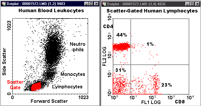
These information is given in page 10 and page 30.
https://www.mre.vghtpe.gov.tw/news/tables/p...w_Cytometry.pdf
What is the role of FSC vs Counts and SSC vs Counts in flow cytometry graphs? What information we can find using these graphs?
I will be very thankfull if you could pass on this information for my future.
Thanks a lot
Regards,
CellSpecific.com on Dec 6 2009, 04:34 PM said:
Thank you very much for your information. I have one more question...
I am planning to perform immunophenotyping soon, I am using both primary and secondary antibodies for this analysis. Can I perform Immunophenotyping without taking PI as counter stain i.e by taking these graphs only.
FSC Vs SSC and FL1 Vs counts.
I will thankful ones again if u can reply for me..
Thanks.
PI is not counter stain. It is stain to exclude dead cells. Those cells that take up the stain are all dead cells and give aberrant readings. So, you cannot be confident about your data unless you are already working with dead cells. Example, when I work with fixed cells for flow cytometry, I do not put PI as there is no meaning of that. However, there are other stains also that can replace PI if U really do not want to have PI.