Confusing bands from PCR - not primer dimer, not product (Nov/06/2009 )
Hi,
I have been getting strange bands on my gel from PCR. They are very clear and they seem too big to be primer dimers. Also, I ran the reaction again reducing the primer concentration from 2.5 µl to 1.5 µl and got the same bands. The product size is 875 bp so I don't think the bands are product either. I am a bit confused.
The agarose gel is 1.5% cast and run in 0.5X TBE buffer, plugged up for 45 minutes.
Please check out the attached picture and let me know what you think.
Thanks very much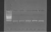
Hi tomato,
Those bands don't look like primer dimers to me, either, but could you please indicate what marker you are using? Knowing what the bands are may help us determine what is wrong.
regards,
lab rat
The marker is P7-43DF3/R1
are u sure your primers are working fine??
i mean is it that u used to get results and now all of a sudden you are not.. or u are doing it for the first time and this result!!!
Sorry, I meant: what DNA ladder are you using? What are the relative molecular weights of the ladder's bands?
The ladder is a Promega 100BP DNA ladder. Smallest band:100, Brightest band:500, Largest band: 1500.
I have not gotten the primers to work before, but I know that it should work.
I ran the experiment again with half the primer concentration. I also ran it on the gel for much longer. The bands were no longer sharp and there appeared to be 2 in each lane. I am going to try playing with the Mg++ and BSA concentrations and further decrease the amount of primer. Good ideas? Any suggestions?
Thanks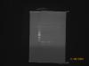
tomatoNC on Nov 9 2009, 03:48 PM said:
I have not gotten the primers to work before, but I know that it should work.
I ran the experiment again with half the primer concentration. I also ran it on the gel for much longer. The bands were no longer sharp and there appeared to be 2 in each lane. I am going to try playing with the Mg++ and BSA concentrations and further decrease the amount of primer. Good ideas? Any suggestions?
Thanks
Hi tomato, I think those are primer dimers and you need to optimise your reaction.
What have you tried so far? And also, what is your exact protocol (concentrations, template, temperatures...)
I'd recommend you try different primer concentrations, different MgCl2 concentration, and if you have a thermocycler that allows you to do gradient try that to find out the best annealing conditions. Also, you might need to try different template concentrations.
Hope this helps, the more detail you give us the more we can help you

I am using a PCR Master Mix that contains (per sample)
5µl 10X buffer
3.2 µl dNTPs
0.2µl Taq 1X
2.5 mM MgCl2
1 µl BSA
I use 11.9 µl of MM with 1µl DNA. In the first picture I was using 2.5µl of each primer and in the second picture I used 1.5 µl of each primer. Then I add enough nuclease free water to make each sample 25µl. Everything is kept on ice. I centrifuge each PCR tube before and after PCR.
The program on the Master Cycler
1) 94 degrees for 3:00 minutes
2) 94 degrees for 30 sec
3) 48 for 30 sec
4) 72 for 2 minutes
Steps 2-4 repeated 31 times
5)72 for 8 minutes
After PCR, 10 µl of each sample is mixed with 2µl loading dye and run on a 1.5% agarose gel in 0.5X TBE at 70 V.
So far, I have decreased the primer amount, increased the primer amount, increased template concentration, increased Taq, and increased dNTPs. The only bands that have showed up yet have been too short and of varying clarity.
Hi Tomato,
I'm just double-checking my understanding of your master mix. Are you adding 5 ul of 10X buffer to a 25 ul reaction? What are the concentrations of your dNTPS?
Thanks,
lab rat
tomatoNC on Nov 9 2009, 05:04 PM said:
5µl 10X buffer
3.2 µl dNTPs
0.2µl Taq 1X
2.5 mM MgCl2
1 µl BSA
I use 11.9 µl of MM with 1µl DNA. In the first picture I was using 2.5µl of each primer and in the second picture I used 1.5 µl of each primer. Then I add enough nuclease free water to make each sample 25µl. Everything is kept on ice. I centrifuge each PCR tube before and after PCR.
The program on the Master Cycler
1) 94 degrees for 3:00 minutes
2) 94 degrees for 30 sec
3) 48 for 30 sec
4) 72 for 2 minutes
Steps 2-4 repeated 31 times
5)72 for 8 minutes
After PCR, 10 µl of each sample is mixed with 2µl loading dye and run on a 1.5% agarose gel in 0.5X TBE at 70 V.
So far, I have decreased the primer amount, increased the primer amount, increased template concentration, increased Taq, and increased dNTPs. The only bands that have showed up yet have been too short and of varying clarity.
What is the actual concentration of your primers? and dNTPs?
I think your annealing temperature is way too low, what are the Tm of your primers?
Also, for a 875bp product 2min is a too long extension, I dont think you need more than 45sec
Finally, following lab rat's question: when you say you use a master mix, do you make this master mix yoursef? if so, are you adding 5ul of 10X buffer to a final 25ul.... ie, 2x final buffer concentration ?
What about the 2.5mM MglC2, is that the final concentration in your sample (ie. in the 25ul) or the concentration in the master mix?