Only borders stained in Silver!!! - (Oct/29/2009 )
Hey all.. I am facing a unique problem.. at least i have never seen such a thing in my life till now!!!
I am working with glycoproteins and one of my glycoprotein stains only at the borders when silver stained!!!
i have only one explanation that being that the protein is more heavily glycosylated than the rest leading to masking of the stain may be??!!!
Wat do you say?? Anyone has seen such a thing??
I will do a coomaaie and see if it stains and post when it is done!!!
Also i forgot to mention that i had run a gradient and a 12% gel both giving me the same result and i ran a NR sample..
If i do a reduced sample run may be it will break the disulphiode bond and change the exposure of glycans and stain better?? or even weorse!!!
let me try this too..
any other suggestions??!!!
are you referring to the borders of the bands or the whole gel (a picture would be nice)?
silver stain may interfere with subsequent analysis. i know you can use a dye-based stain (eg coomassie) after silver but reactive stains may be affected.
mdfenko on Oct 29 2009, 09:15 PM said:
silver stain may interfere with subsequent analysis. i know you can use a dye-based stain (eg coomassie) after silver but reactive stains may be affected.
hi md.. i m referring to the band.. not the gel border!! will attach the gel pic as soon as i can.. right now scanner not workin!!!
i will destain the silver stained gel.. then coommaassiiiiee it.. if it gets stained, i l have to think more... if it does not too.. then i will reduce and run the sample!!!
hi md.. here you go.. the gel pic.. and ya i also cant explain why the artifact (the straight line thru the gel) is persent in both the gels exactly at the same position... one is 12% and the other is gradient!!! you d know which one is wat 
both were run in the same unit may be some thing in the run buffer... any other erason o observations.. do let me know!! and the origical querry still stays.. the title!!!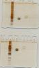
nice pictures.
it looks like you have the halo effect on both samples, just more prominent on one than the other. this may be due to sample load. have you tried less sample?
is this sds-page? i ask because you said, earlier, that you didn't reduce your sample. why not?
the band across the gel can be an artifact caused by the presence of keratins (from dust) in one or more solutions or it can be caused by a trailing ion from the electrode buffer. it may not show up with coomassie stain (silver is a lot more sensitive).
are you going to do the glycoprotein stain in the gel or are you going to transfer to a membrane prior to blotting?
halo.... can u elaborate on that effect please??
ya i did a coomassie and dint get that horizontal artifact as you said.. so that must be some thing in the buffer or the keratin as u say!!!
i have loaded just 3 microgram so oerloading cant be the reason i think!!!
i dint reduce cause i was interested in some high moleular weight bands which i anyways dint get 
but i am not going to blot it... but subssequently sometime i have to do that...
my worry is if this is due to the glycan which is masking the stain to interact with the protein, it might effect the epitope too and my antibody also might not work!!! or might have to use a very low dilution!!!
nice pictures??!!! was it a taunt!!! 
Pradeep Iyer on Oct 30 2009, 11:41 AM said:
ya i did a coomassie and dint get that horizontal artifact as you said.. so that must be some thing in the buffer or the keratin as u say!!!
i have loaded just 3 microgram so overloading cant be the reason i think!!!
i dint reduce cause i was interested in some high molecular weight bands which i anyways dint get
3ug of a purified protein is a large load on a minigel. the bands are pretty big, we usually want flat, thin bands. did you load a large volume?
also, in what buffer is the sample (besides the sample loading buffer)? buffer components can cause all kinds of grief.
my worry is if this is due to the glycan which is masking the stain to interact with the protein, it might effect the epitope too and my antibody also might not work!!! or might have to use a very low dilution!!!
i wasn't referring to immunoblot. we have performed glycoprotein staining both in-gel and on-membrane.
not at all. it shows what we need to see.