about measuring luciferase activity in living cells - (Sep/10/2009 )
Hi all:
Dose anyone have the experience to measure luciferase activity from living cells rather than cell lysates? If so, could you please give me some suggestions?
Thanks in advance!
--Stormlina
As far as i am aware you always need to lyse the cells to measure the luciferase activity.
there are various products available for measuring which all seem to have differing activities in terms of light intensities and half life time, we have always found promega to be quite good in this regard, oneglo i think is a good product
we have also used antibodies against luciferase, also available from promega (and others) to immunostain and western blot for the presence of luciferase in transfected cells, but again this requires fixing and lysing the cells.
Stormlina,
It is technically possible to measure luciferase activity in intact cells by disolving luciferin into your culture media, letting it equilibrate for 1-2 hours, then reading. While this will give a signal, I warn you that it is not a very reliable method as the reading will be a small fraction of the amount obtained by measuring luciferaes in a lysate with a quality luciferase assay reagent (and I'm glad to hear cotchy finds our products to work well). The reasons for this include temperture effects on the luciferase as you move the sample from incubator to the reader, but more importantly the dependancy on ATP to generate light. The ATP in the cell will not be saturating, so the light output will be dependent not only on the luciferase expression level but also on the ATP concentration. Luciferase assay reagents are designed to keep all reaction components at an optimal concentration to allow quantitation of the lucferase enzyme.
You may note that many people performing cell engraftment study with luciferase-expressing cells show live cell lucifersae data. In these cases the absolute luciferase expression level is not critical and the luciferase is highly expressed to simly label living cells to be monitored in animals. In these cases the live cell method is probably acceptable.
Hopet his helps. Please let me know if I can be of any assistance.
Regards,
Kevin
Hi Kevin:
Thank you very much for the nice information! I am planning to measure luciferase signal in living cells as well as in animal later on. I read some paper talking about measure in vivo and no extra ATP is necessary. I am wondering whether you have any idea that if I am going to measure the signal in in vitro cultured cells, do I need to add some ATP besides luciferin?
Thanks.
Stormlina
KevinK on Sep 11 2009, 08:34 AM said:
It is technically possible to measure luciferase activity in intact cells by disolving luciferin into your culture media, letting it equilibrate for 1-2 hours, then reading. While this will give a signal, I warn you that it is not a very reliable method as the reading will be a small fraction of the amount obtained by measuring luciferaes in a lysate with a quality luciferase assay reagent (and I'm glad to hear cotchy finds our products to work well). The reasons for this include temperture effects on the luciferase as you move the sample from incubator to the reader, but more importantly the dependancy on ATP to generate light. The ATP in the cell will not be saturating, so the light output will be dependent not only on the luciferase expression level but also on the ATP concentration. Luciferase assay reagents are designed to keep all reaction components at an optimal concentration to allow quantitation of the lucferase enzyme.
You may note that many people performing cell engraftment study with luciferase-expressing cells show live cell lucifersae data. In these cases the absolute luciferase expression level is not critical and the luciferase is highly expressed to simly label living cells to be monitored in animals. In these cases the live cell method is probably acceptable.
Hopet his helps. Please let me know if I can be of any assistance.
Regards,
Kevin
What cells are you examining for luciferase activity?
prostate cancer cell CWR22-RV stably transfected with luciferase gene.
Thanks.
eberthella on Sep 17 2009, 04:30 AM said:
Stomlina,
No, it is not necessary to supplement any ATP. In your case, the luciferase is a marker for the cells and intensity may vary based on their viability (as ATP is only present in live cells and ATP and luciferase would degrade rapidly upon cell death). The ATP concntration is usually only an issue when using luciferase to quantitively measure an activity that regualtes luciferase expression.
Hope this helps, and good luck in your studies..
Kevin
Is it Firefly or renilla luciferase? As far as I know, Promega offers substrates for in vitro Renilla luciferase measurement in living cells (Enduren or viviren I think, can't remeber exactely)
Stardust
PS: maybe they have something for firefly as well check on their website.
Thanks for the excellent point, Stardust. My comments related to firefly luciferase. Promega does indeed provide Renilla luciferase substrates for use in live cells. These products, EnduRen and Viviren, are derivatives of the typical coelenterazine substrate typically used by Renilla and offer decreased background and in the case of EnduRen, a very long lived signal.
Kevin
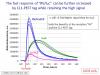
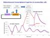
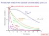
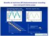
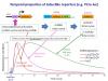
By the way, people who have an appropriate real-time recording luminometer/"Lumicycler" (that keeps plates at 37°C) can benefit from getting a continuous recordings from live cells - without any lysis (or by adding it at the final stage as another control).
This way you can add (e.g. 0.1 mM or more) firefly luciferin to the medium and record for several days.
Each well/plate can serve as its own control before the treatment/induction and then provide a continuous bioluminescence curve (instead of discrete points obtained by lysis of different wells/plates).
This approach is widely used in the field of circadian gene expression and beyond.
Some illustrative slides: