uggly, non-focus and diffuse bands in my Western.. - Can you see my image of my Western to LC3? (Aug/22/2009 )
Hi.
Recently I run a Glycine-SDS-PAGE to detect the autophagic protein LC3-I and II (16 and 18Kd) . I loaded the recommended quantity of sample: 40ug. The transfer was OK, but my bands appears diffuse with a long shadow. I run to 80V until the limit between gels, and then to 120V, 3hrs in total. I prepared new and fresh buffers to make the gels, but nothing..I get the same result.
Which other aspect-change-mistake-trick should I consider?
If you need to see my recent Western Blot to estimate what might happen (like a doctor), I have attached the image in this message.
Thanks in advance.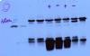
It's not too bad as far as westerns go. Perhaps try loading a little less protein, use a stronger blocking solution, or more stringent wash conditions to cut down on some of the non-specific binding.