J774 and Flow cytometry....are they really big cells ? - (May/17/2012 )
Kaixo,
I've been playing with flow cytometry and some J774 cells for a while but am a little confused by the FSC v SSC plots I'm seeing. I would really appreaciate it if anyone who uses these cells for flow can give me some advice!
I have experience growing and doing cytotoxicity and various other assays with J774 cells and I realise that they are extreamly sticky and grow like made! To reduce their grown I have reduced the FBS to 5%, and to unstick them I have found the best way is to pipette (w/p1000) and re-use the flasks for <10 passages as they seem to detach better when their home 'isn't new', as such 
I have run them on the flow cytometer with PBS, with PBS+BSA, with media; after scrapping, after pipetting, after using Sigma cell uptake solution; after fixing and live .... regardless I always seem to get 2 populations of cells (see attachment), one that I have called 'P1' (red), and then the other stuff which looks like debris.
I guess my cells are 'P1', but is it normal for them to be so big ? (my FSC voltage is 4, and the max. is 1000!!), and I only just fit them onto the plot. And why so much debris ? Is this normal for J774s ? I realise I can set my FSC threshold higher (for example 1000 rather than 200) and then this would "remove" my debris, albeit only visually, but is it normal ?
Any advice would be greatly appreciated !!
Eskerrik asko, Moo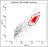
You should use FSC-H or -W instead of -A (what is the difference?), then your cells will fit on the plot. You'll need a FSC voltage of 40 to 70 to get them into the middle. Run a tap water sample (cell-free) and find that the dots left of your cell population are in fact bacteria, which are always present everywhere. I suggest you set the FSC threshold to ~50,000, otherwise your quantification will be screwed (events include bacteria or not?).
I would have thought the red population was doublet/triplet etc. cells while the rest were singlet cells.
Rsm on Fri Jun 1 09:58:15 2012 said:
Run a tap water sample (cell-free) and find that the dots left of your cell population are in fact bacteria, which are always present everywhere
Bacteria? I wouldn't have expected that many, if it were in fact bacterial contamination. But what would be the source anyway?
I've never thought about possible bacteria in my flow. I guess it depends what you use, but I make all of my FACS solutions using PBS or Baxter water, both of which are sterile. But if you gate it out and set your stopping gate on your desired population only, it won't affect your analysis anyway.
Could you look at a portion of a sample under the microscope to check if it is the doublet/triplet thing bob1 mentioned? Seems like a reasonable explanation if your cells are so sticky. Or what about passing your samples through some gauze first, to filter out anything cell clumps etc.
Does seem like a pretty tight population though... so maybe what you are seeing is just debris?
Have you tried using a live/dead discriminator stain? That could help you figure out what is cells and what is junk?
bob1 on Sat Jun 2 02:02:37 2012 said:
I would have thought the red population was doublet/triplet etc. cells while the rest were singlet cells.
Then he would have quite many cell doublets, and very few single cells. Even if he fixed his cells this amount of doublets is unlikely.
leelee on Sat Jun 2 06:34:48 2012 said:
Rsm on Fri Jun 1 09:58:15 2012 said:
Run a tap water sample (cell-free) and find that the dots left of your cell population are in fact bacteria, which are always present everywhere
Bacteria? I wouldn't have expected that many, if it were in fact bacterial contamination. But what would be the source anyway?
I've never thought about possible bacteria in my flow. I guess it depends what you use, but I make all of my FACS solutions using PBS or Baxter water, both of which are sterile. But if you gate it out and set your stopping gate on your desired population only, it won't affect your analysis anyway.
Could you look at a portion of a sample under the microscope to check if it is the doublet/triplet thing bob1 mentioned? Seems like a reasonable explanation if your cells are so sticky. Or what about passing your samples through some gauze first, to filter out anything cell clumps etc.
Does seem like a pretty tight population though... so maybe what you are seeing is just debris?
Have you tried using a live/dead discriminator stain? That could help you figure out what is cells and what is junk?
I think it appears to be "many", because it's not a density plot nor does he provide numbers. I guess that population is between 0.5 to 1%, that's not that much. There's certainly a decent amount of cell debris as well, not only bacteria. Sources of bacteria are everywhere, us, the hood, FBS, pipets... you get the point. Remember that most antibiotics don't kill bacteria, so even if there's no obvious contamination, it doesn't mean that it's still sterile. Try running some sterile FACS buffer alone, I bet that you'll get some events.
leelee, are you using FSC-A or H? Does your plot looks similar when you use FSC-A? At least mine does. Microscope and live/dead are good ideas, but changing the scale may be easier and quicker.
I disagree that 0.5- 1% of a cell sample being bacterial contamination is not very much. That is huge, in my opinion. That means that for every 99 cells that are what you want, you would have 1 bacteria. If that were truly the case, and this was bacteria, my advice would be to throw it out and start again. You will never be able to be sure of your results if you have contamination present. Who knows what they could be doing to the cells' behaviour.
I don't use antibiotics to culture my cells, so any bacterial contamination (even if it were low level to begin with) would show up very quickly.
There is definitely no bacteria in my FBS! It comes to us sterile from the source, and any contamination would be very quick to show up in such a nutrient rich liquid. I've seen a contaminated vial of FBS become turbid with bacteria in a matter of days at 4C.
I agree that bacteria are everywhere, but if you use correct aseptic technique and sterile solutions, you won't get any in your cell cultures or your solutions.
I use FSC-A, and to be honest haven't ever looked at the other two (FSC-H or FSC-W) but I can't imagine there would be a huge difference, as they all measure slightly different variations of the same thing- unless I've had it wrong all this time...
I think it is important for moo to be confident that what they are gating on is single cells- so having a quick look with the microscope will take only minutes and give moo piece of mind.
The live/dead thing is probably worth doing at some point too- it is a good thing to know, how many of your cells that you are analysing are alive or not.
Perhaps, moo, you could consider contacting technical support from your instrument's manufacturer. They may be able to help you out? Or finding someone nearby who is experienced on this machine and knows how to help you?
Good luck 
I agree, my initial statement was a bit misleading. My proposal to measure a water sample to see bacteria was rather to get an idea of the size of events that he measures. Bacteria are present in tap water and will be at the same size of the events he observes. However, I still wonder why leelee disagrees with my statement that he has cell debris in his preparation? How do you prevent that, leelee?
If an event passes the laser beam a Gaussian curve is produced in the detector (signal intensity vs time). FSC-A is the area under the curve, while W is the width and H is the heigth. They relate approx. A=H*W (way more complicated in fact), so there is in fact a huge difference. W being the smallest, H bigger and A the product of the two.
Rsm on Sun Jun 3 12:39:14 2012 said:
However, I still wonder why leelee disagrees with my statement that he has cell debris in his preparation? How do you prevent that, leelee?
I don't disagree that there will be cell debris in the preparation, I never said that? I apologise if that was unclear. I actually DO think that it is most probable that what we are seeing on the plot is debris.
And as for the FSC, I suppose I wrote that rather poorly too. I meant that they are using the FSC to assess the signal from the same thing/particle in different ways, rather than any comment on the values themselves.
No worries... I wonder if our basque friend is still around or if we chased him away with our academic discussions?
haha I was wondering the same thing